Dental Imaging Means?
Dental radiographs, also known as X-rays, are radiographs that are used to diagnose cavities, bone loss, hidden dental structures, and benign or malignant masses.
A controlled burst of X-ray radiation penetrates oral structures at various levels based on varying anatomical densities.
Because less radiation reaches the film through the teeth, they appear lighter.
Because X-rays can easily penetrate these less dense structures, dental caries, infections, and other changes in the bone density, as well as the periodontal ligament, appear darker.
For more detailed information on this and to get this treatment, you must visit Park dental clinic which is the best dental clinic in Dwarka sec7.
Procedure of Dental Imaging
- Depending on the density of the material, dental restorations may appear lighter
or darker. - A dental patient typically receives a small dose of X-ray radiation which is comparable to the dose received during a cross-country aeroplane flight or a few days’ worth of background environmental radiation exposure.
- Utilizing a lead shield, lead apron, and sometimes a lead thyroid collar further reduces incidental exposure.
- When the X-ray source is activated, the technician’s exposure is reduced by stepping outside the room or hiding behind sufficient shielding.
- Because photographic films are sensitive to natural light, they must be developed after being exposed to X-ray radiation.
- This process typically involves exposing the film to a series of chemicals in a dark room.
- The above process may take a long time, and the patient may be exposed to additional radiation if retakes are required due to incorrect exposures or mistakes made during the development process.

Future Aspects
As technology advances, digital X-rays, which replace film with an electronic sensor, are becoming increasingly utilized in dentistry to address some of these issues.
They may require less radiation and are processed much faster than traditional radiographic films, and they can frequently be viewed immediately on a computer.
Digital sensors, on the other hand, are very expensive and have historically had poor resolution, but this is much better with modern sensors
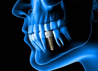
When is Dental Imaging required
- Root canal
- Dental implants
- Orthodontics
- Oral surgery.
Those are some of the factors when this process is needed, but before taking a decision you should consult your dentist and Dr. Neetu Singh at Park Dental Clinic in Dwarka sec7 is the best dental clinic available.
Benefits of Dental Imaging:
- Technology is safe for pregnant women because it produces less radiation than conventional x-rays.
- When compared to conventional x-rays, the procedure takes significantly less time. Most of the time, it only takes 20 seconds.
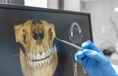
- 3D images are better at showing your teeth, skull, and jaw because they are more focused.
- Demonstrate the differences between the tissues in your mouth, making it simpler to treat dental issues and diagnose them.
- The dentist can examine the quality of your bone tissue with the assistance of 3D imaging, which can be crucial during treatment.
- With 3D imaging, the dentist can show the patient the images and explain them to them. He can magnify and comment on the images to help you understand them better.
- The machines are simple to use for any trained practitioner thanks to the user-friendly cone beam technology.
- The cost of 3D imaging is relatively low and there are no aftercare instructions, and the procedure is comfortable.

Dental Imaging Safety
- According to NRC guidelines, adults can be safely exposed to 5,000 millirems of radiation annually.
- 500 millirems of radiation can be safely given to children every year.
- A 60-year-old person could have safely been exposed to 60,000 millirems of radiation over the course of her life.
- Over time, individuals should not be exposed to more than 1,000 millirems of radiation times their age. Dental x-rays fall far short of this safe limit.
- Dental x-rays have the lowest radiation doses from these medical procedures, but medical x-rays account for the majority of man-made radiation exposure each year.
- The NRC estimates that a full-body CT scan will produce 1,000 millirems of radiation.
- The dose of a dental x-ray is only 1.5 millirems. In light of the fact that food contains an average of 30 millirems of radiation, x-rays only expose patients to trace amounts.
- Although x-rays are one of the safest medical procedures, none are completely risk-free. To further reduce risk, many dentists now use digital x-rays rather than film.
Best dental clinic to visit
If you are searching for a dental clinic which gives the best service, treatment and also is hygienic then you must visit Park dental clinic without giving a second thought, because it is the best dental clinic in Dwarka sec7 and Dr Neetu Singh over there is the best dentist. So for all of your dental needs, Park Dental Clinic should be your first choice because they use cutting-edge technology and have specialized expertise for Root canal, Ortho and dental implant treatment.
Visit Park Dental Clinic right away!
References
https://pubmed.ncbi.nlm.nih.gov/29913523/
https://en.wikipedia.org/wiki/Dental_radiography
https://www.dentalcare.com/en-us/ce-courses/ce512/digital-imaging-basics
https://www.healthline.com/health/dental-x-rays
https://dentalhealthsociety.com/general/are-dental-x-rays-safe/